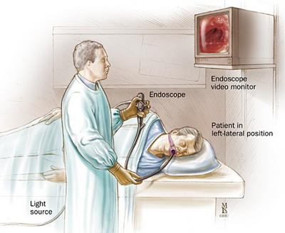
EHD or esophagoscopy is an advanced endoscopic technique that examines the upper part of the digestive tract all the way down to the intraluminal lymph nodes. Esophagoscopy is usually performed on patients who have no other means of diagnosing gallstones or are unaware of their presence.
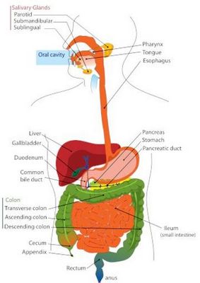
The esophagus consists of a tubular structure called a lumen, which is a passageway for food and fluids and is lined with epithelium. At the end of this tube are two types of small and large intestine. The larger intestine is called the descending colon or ileum, and the small intestine is called the ascending colon or descending spleen.
The small intestine is located just below the stomach and is connected to it by a rigid duct. The large intestine is located directly under the stomach and is associated with it by the peristaltic movement of stomach acids from the stomach and duodenum to the small intestine. As you can see, the small intestine is much smaller than the large intestine. During an esophagoscopy, your doctor will take a sample of your blood to test for gallstones.
A catheter inserted into the esophagus is called an endoscope. It passes through the esophagus into the duodenum to remove material. The endoscope uses two thin tubes – one to inject a dye solution and the other to insert a liquid-filled catheter. A needle is used to inject the dye solution through a catheter into the esophagus.
If you experience unusual discomfort in the lower abdomen or pain in the small intestine, see your doctor immediately. This could be a sign of gallstones. You may need to have gallbladder surgery if any.
Ego use is described as an advanced technique. Since it is an endoscopic method, it simplifies diagnosis and does not require the use of an anoscope or X-rays. The small intestine and pancreas are often visible with an endoscope.
Egd can be used to diagnose gallstones, polyps, bile duct obstruction, cystic fibrosis and carcinoma, and even some cancers. It has been found to be helpful in treating esophageal reflux disease and gallstones. The end is now widely used in the United States.

EHD is considered a safe procedure and does not cause serious complications. The procedure is relatively simple and generally safe.
After the dye is injected, the dye is injected into the small intestine through a rigid duct. The dye is then absorbed into the tissues. Some cells in the small intestine do not die when injected. However, some will absorb the dye. The remaining cells absorb the dye into the bloodstream as absorption takes place.
After the dye is absorbed into the small intestine, the doctor examines the color of the cells. If the color of the cells is darker than the color of the environment in the small intestine, then gallstones can be identified at the end. and the catheter is removed to retrieve them. for analysis.
If the dye is found in the urine, it will indicate the presence of gallstones and that a catheter has been inserted into the small intestine. and the gallbladder is removed. If there is no gallbladder, a catheter is inserted into the duodenum and a dye is injected. During this time, a catheter is used to inject dye into the catheter to empty the gallbladder.
The catheter is then removed and the catheter removed. If there are no gallstones, the procedure will end.
Egd is a relatively simple procedure and the risk of complications is very low. However, it is not recommended for people who are not well trained or inexperienced.